
Graphical Abstracts and Highlights
What is a graphical abstract?
A graphical abstract (GA) is a stand-alone image summarizing the major findings of your Original Article in the form of a pictorial take-home message. GA are required only Original Articles. It should be submitted together with three bullet points (Highlights) that summarize the main findings presented in the paper.
What is the aim of preparing a graphical abstract?
GAs are designed to draw attention to the target audience, serving as a quick graphical summary of the research findings.
1. Graphical abstract – image:
Please create your GA image in PowerPoint using the Allergy GA Template and graphics from the Allergy Graphics Collection. You can simply copy and paste the relevant shapes/figures to your GA.
2. Highlights - text
-
Include: title of the manuscript, summary of the study in 3 bullet points, spelled-out abbreviations used in GA and highlights in alphabetical order
-
Word limit for bullet points: 60 words in total
-
File Format: doc, docx
The Graphical Abstract Image Should
-
be different from the figures included in the manuscript
-
be easy to understand and well-structured
-
be based on graphical elements
-
focus on the main findings
-
contain a minimal amount of text
-
contain simple labels
-
use the available space efficiently without leaving empty spots
-
use the colours from Allergy's colour palette
-
effectively use accent colours to direct the attention to the point of interest
-
correspond with the Highlights
-
only use the background colour HEX #AEBDE9, as in the Allergy´s graphical abstract template
The Graphical Abstract Image Should Not
-
contain a title
-
include any microscope images, CT or X-rays scans,immunoblots, etc.
-
contain light colours on a light background (e.g., yellow on white)
-
contain highly-saturated colours
-
include any graphics downloaded from online resources without obtaining copyrights
-
have white, black or very dark background
-
#000080 (RGB: 0/0/128) → fonts, arrows, frames
-
#AEBDE9 (RGB: 174/189/233) → background darker
-
#DBE6FF (RGB: 219/230/255) → background lighter
-
#FFFFFF (RGB: 255/255/255) → textboxes
-
#CB012D (RGB: 203/1/45) → inhibition arrows
-
#FF2F92 (RGB: 255/47/146) → bar graphs, data points
-
#008F00 (RGB: 0/143/0) → bar graphs, data points
-
#00B0F0 (RGB: 0/176/246) → bar graphs, data points
-
#FFC000 (RGB: 255/192/0) → bar graphs, data points
The graphical abstract image should be submitted together with highlights as separate files by selecting either “Graphical Abstract Image" or “Graphical Abstract Text" from the file designation list in the manuscript submission system. Files should be named as followed:
-
GA_ last name of the 1st author _ALL-xxxx_xxxxxx (saved as JPG, PNG, PDF)
-
Highlights_ last name of the 1st author_ALL-xxxx_xxxxxx (saved as doc or docx)
Examples of Graphical Abstract
Authors are encouraged to follow the examples of good graphical abstracts, prepared according to the guidelines, both in terms of visual aspect and conveyance of the article's take-home message. See more in the graphical abstract archive
-
We assessed whether the prevalence of allergic respiratory disease has continued to increase in the current millennium among Danish adults.
-
Both specific IgE (sIgE) and skin-prick test (SPT) sensitization showed an increase from 2006 to 2013-2016.
-
These trends were supported by a nationwide increase in annual sales (including over-the-counter drugs) of intranasal corticosteroids and oral antihistamines from 1997-2015.
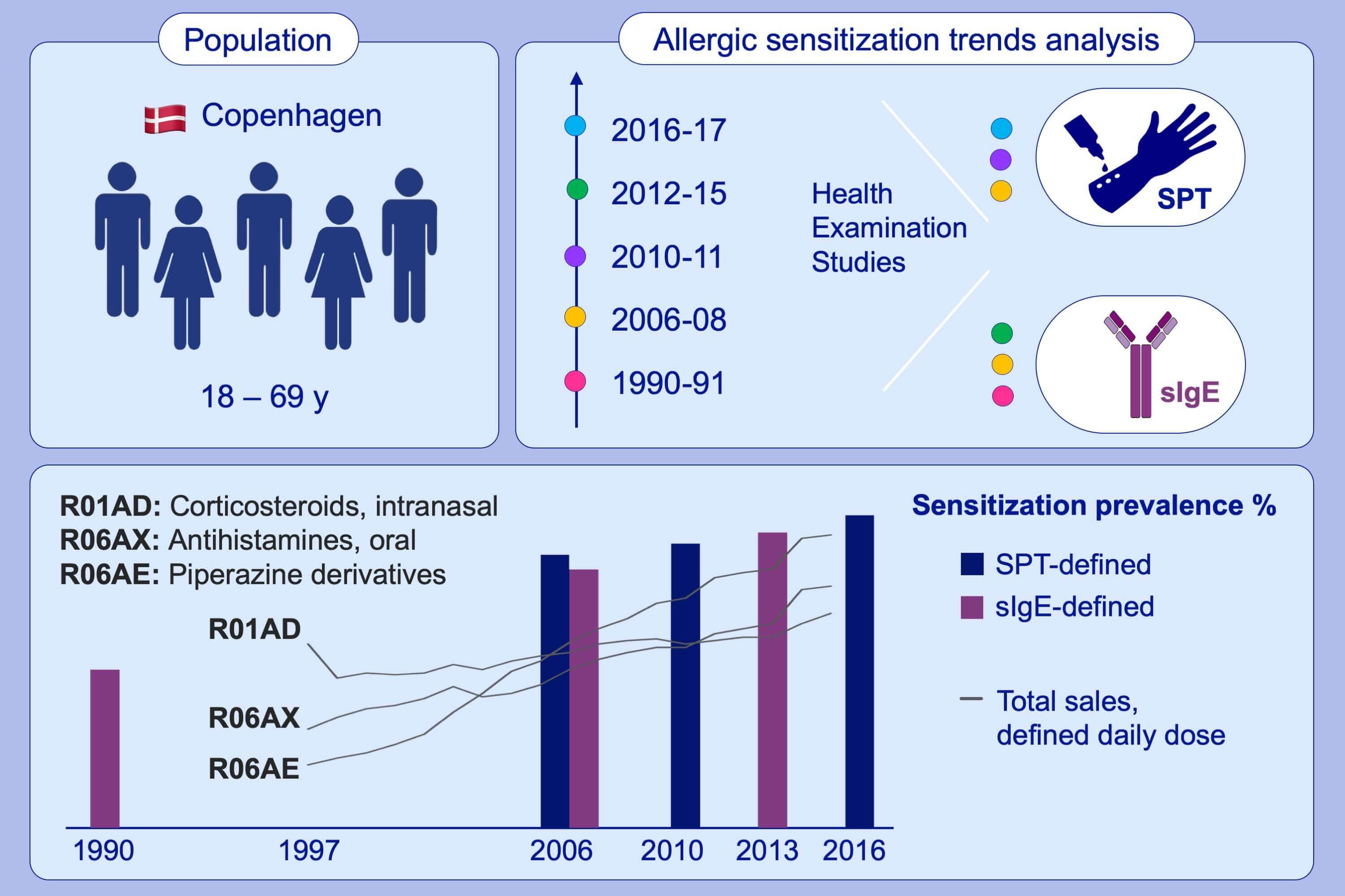
Allergic rhinitis and allergic sensitisation are still increasing among Danish adults
Katja Biering Leth-Møller, Tea Skaaby, Allan Linneberg
Abbreviations: sIgE, specific IgE; SPT, skin prick test
-
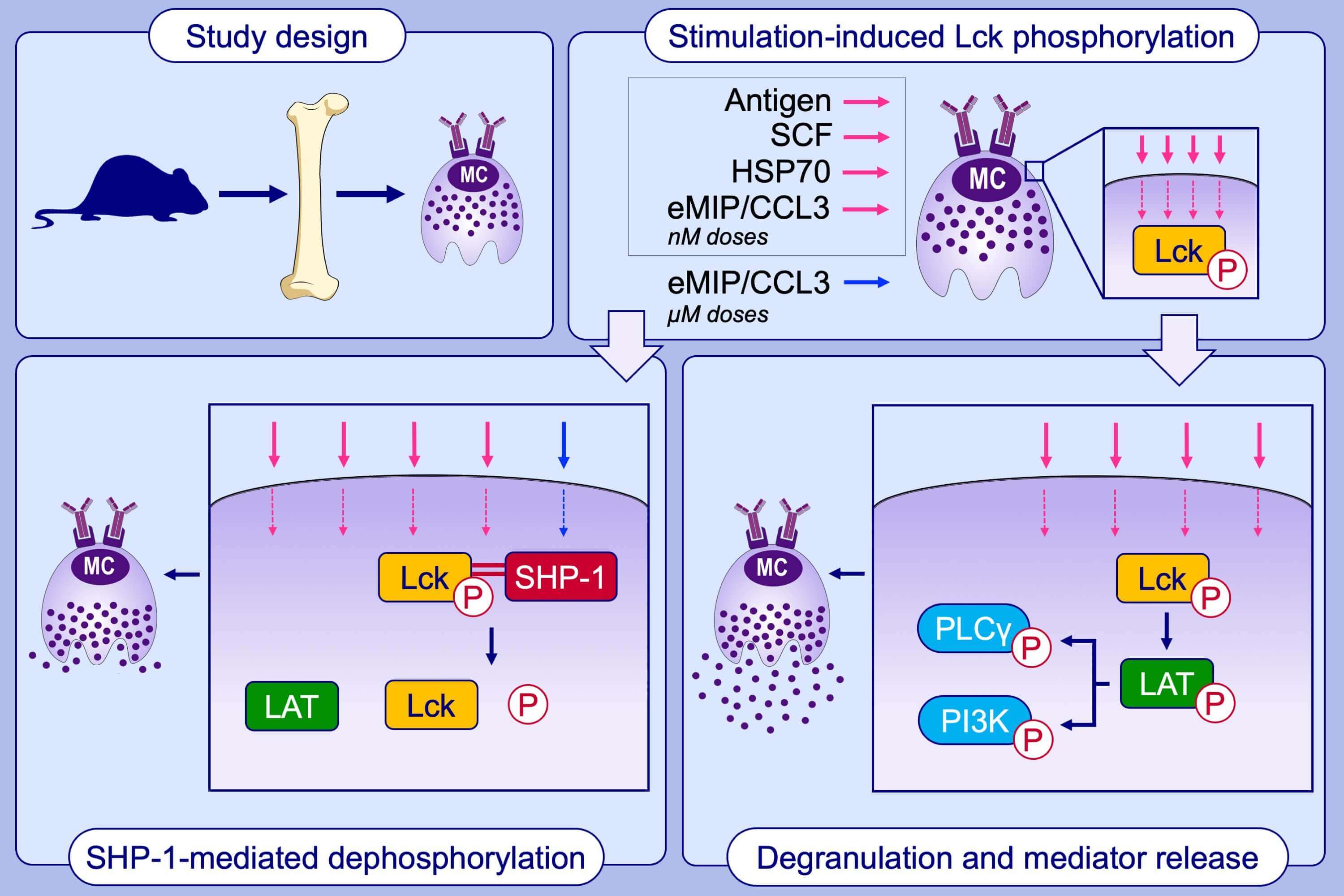
Binding of IgE/Ag, SCF, HSP70, and eMIP/CCL3 to their receptors induces Lck phosphorylation, which in turn phosphorylates LAT.
-
PhosphoLAT triggers phosphorylation of PLC, PI3K, and downstream signaling proteins leading to degranulation and mediator release.
-
All receptors are internalized with the aid of phospho-Lck. Activated by micromolar doses of eMIP/CCL3 or by A770041, SHP-1 dephosphorylates phospho-Lck, whereby receptor recycling, degranulation and mediator release are terminated.
-
Activated by micromolar doses of eMIP/CCL3 or by A770041, SHP-1 dephosphorylates phospho-Lck, whereby receptor recycling, degranulation and mediator release are terminated.
A common signaling pathway leading to degranulation in mast cells and its regulation by CCR1-ligand
Hyeun Wook Chang et al.
Abbreviations: A770041, a selective inhibitor of Lck; CCL3, C-C motif chemokine ligand 3; eMIP, a 69- amino acid variant of human CCL3; HSP70, heat shock protein 70; Lck, lymphocyte-specific protein tyrosine kinase; LAT, linker for activation of T cells; PI3K: phosphatidylinositol 3-kinase; PLCγ: phospholipase Cγ; SCF; stem cell factor; SHP-1: Src homology region 2 domain-containing phosphatase-1
-
Among 1831 studied AD patients, 86.1% were treated with dupilumab.
-
Patients continuing therapy experienced a marked reduction of disease severity during pandemic.
-
The causes of treatment interruption included: fear of increased susceptibility to SARS-CoV-2 infection (24.8%), occurrence of comorbidities (5.9%), age above 60 years (5.2%), SARS-CoV-2 infection (2.8%), close contact with SARS-CoV-2- positive subject (2.4%), other reasons, for example, inability to maintain drug supply, non-medical/unspecified causes (58.7%).
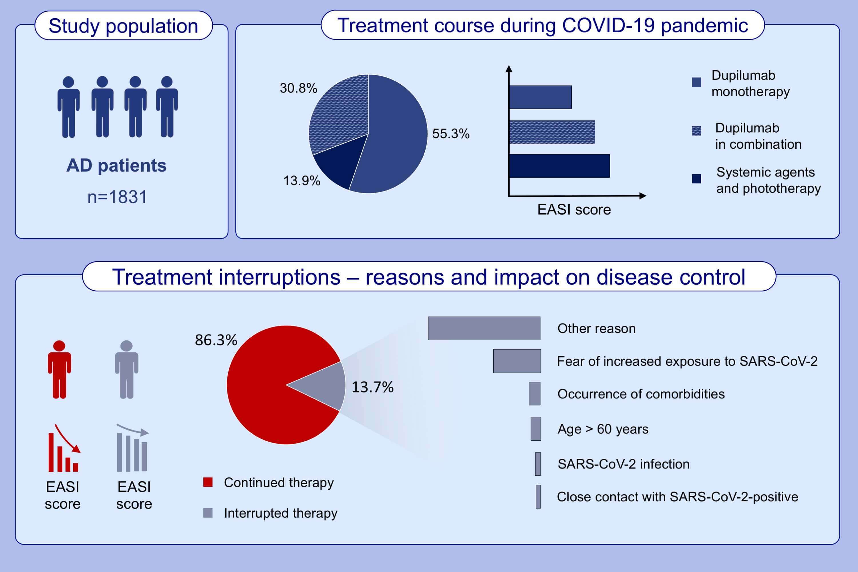
Management of patients with atopic dermatitis undergoing systemic therapy during COVID-19 pandemic in Italy: Data from the DA-COVID-19 registry
Andrea Chiricozzi et al.
Abbreviations: AD, atopic dermatitis; COVID-19: coronavirus disease 2019; EASI, eczema area severity index; SARS-CoV-2, severe acute respiratory syndrome coronavirus type 2
-
This study compares the clinical, radiological, and laboratory characteristicsand longitudinal variations in laboratory parameters of the 289 hospitalized patients with COVID-19 with different severity and clinical outcomes.
-
We propose that elder age, a greater number of affected lobe(s), elevated C-CRP level and increased prevalence of chest tightness/dyspnea and smoking history may be associated with the fatal outcome of patients with severe COVID-19.
-
We identify strong correlations between age, affected lobe numbers, and laboratory variables.
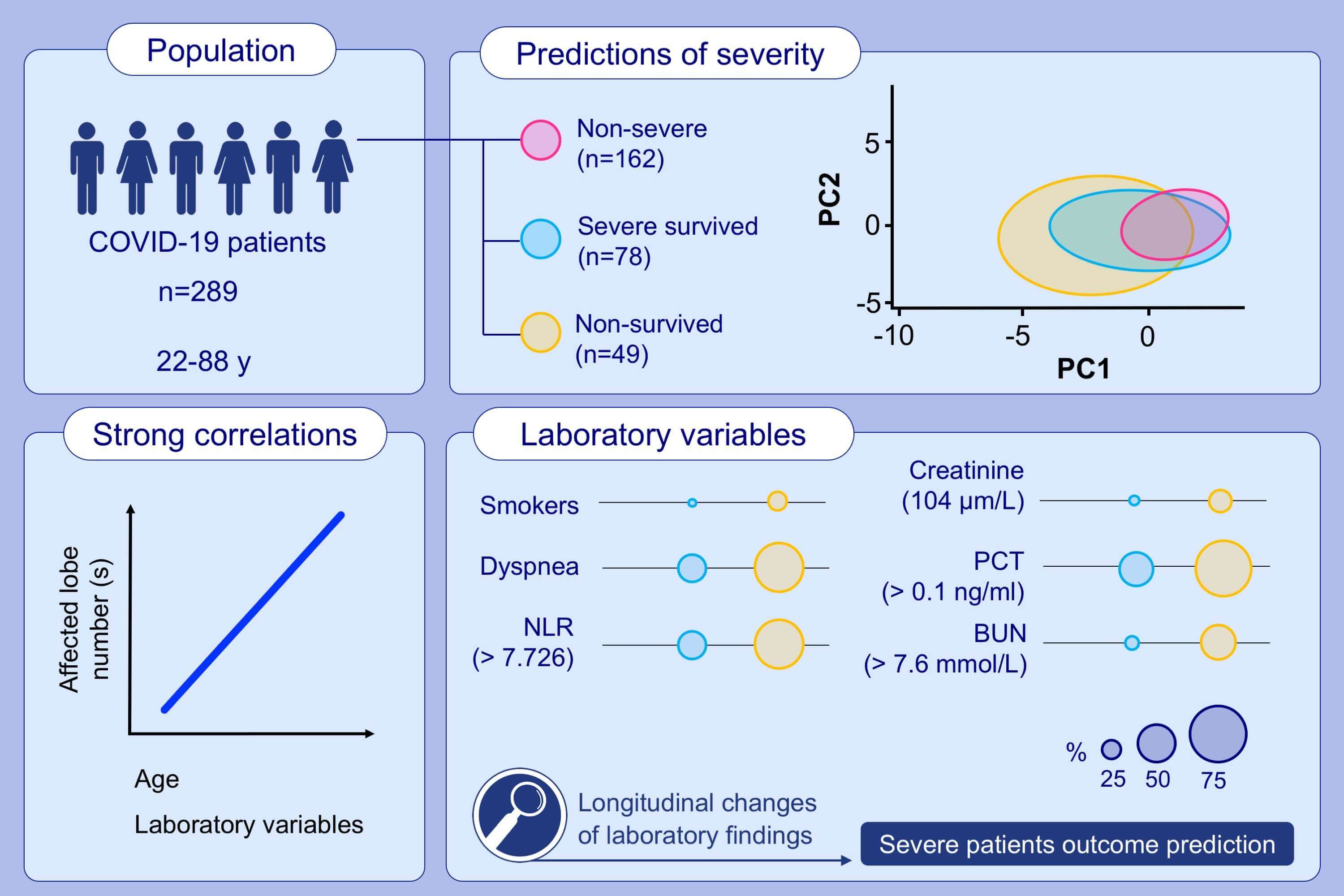
Clinical, radiological, and laboratory characteristics and risk factors for severity and mortality of 289 hospitalized COVID-19 patients
Jin-jin Zhang et al.
Abbreviations: BUN, blood urea nitrogen; COVID-19, coronavirus infectious disease 2019; CRP, C-reactive protein; NLR, neutrophil to leucocyte ratio; PC1/2, principal component 1/2; PCT, procalcitonin
-
Increased sputum and serum TNF markers are associated with neutrophilic asthma, severe asthma and frequent exacerbation history.
-
Azithromycin treatment reduces airway and systemic TNF markers, suggesting an anti-inflammatory mechanism.
-
Azithromycin effect on TNF markers is most pronounced in non-eosinophilic asthma. Effect on serum but not sputum markers is related to sputum bacteria-positive culture, linking antibiotic effect to modulation of systemic inflammation.
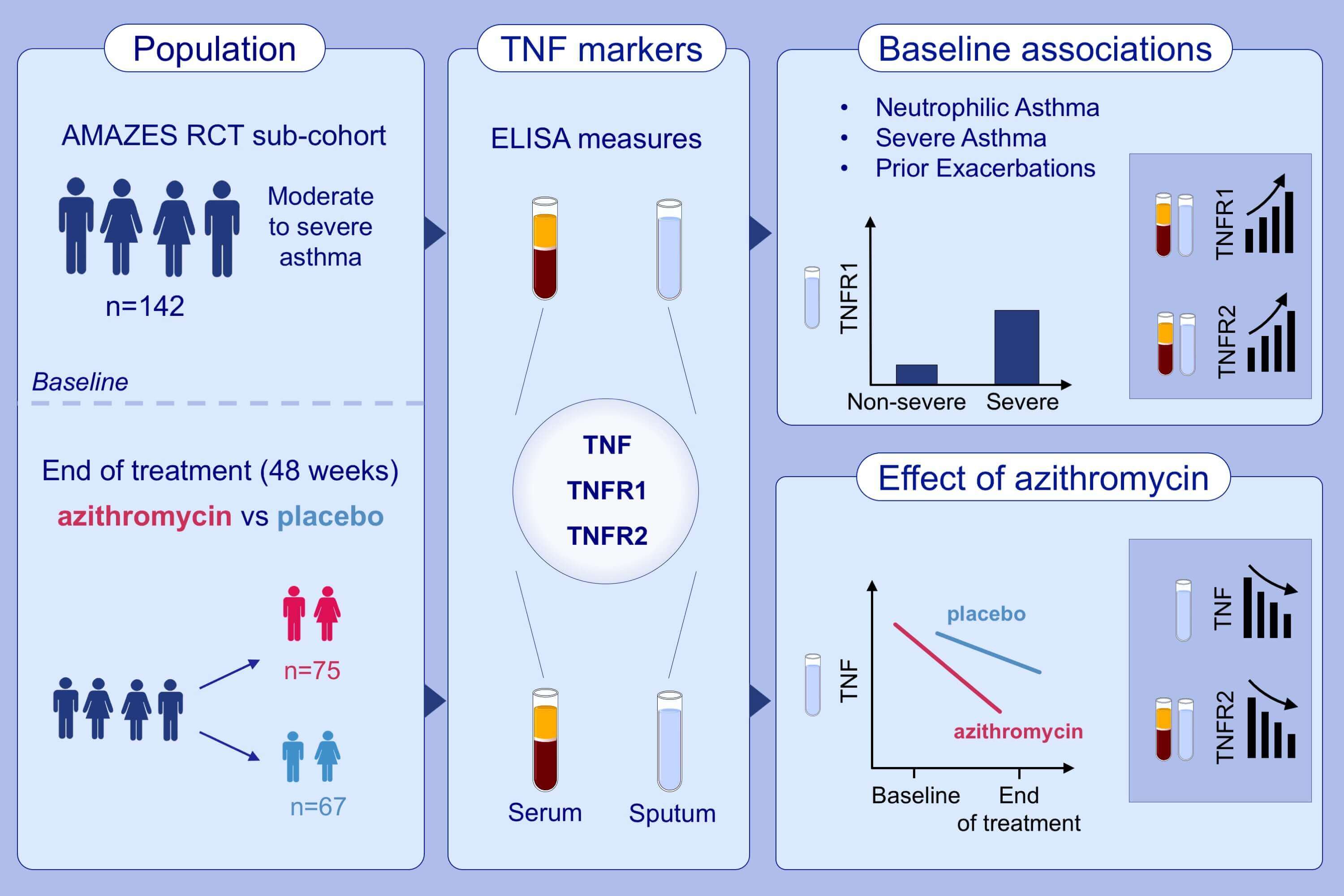
Sputum TNF markers are increased in neutrophilic and severe asthma and are reduced by azithromycin treatment
Natalie M. Niessen et al.
Abbreviations: AMAZES-RCT, Asthma and Macrolides: The Azithromycin Efficacy and Safety study – Randomized Control Trial; TNF, tumor necrosis factor; TNFR, TNF receptor
-
EIS differentiates between AD patients and controls.
-
EIS can characterize lesional and non-lesional skin of patients. During hospitalization, lesions showed a significant increase in EIS that correlated with healing.
-
EIS scores showed a significant inverse correlation with serum biomarkers associated with inflammatory response, particularly chemokines such as CCL13, CCL3, CCL7, CXCL8, and other molecules such as IRAK1, IRAK4 and FG2.
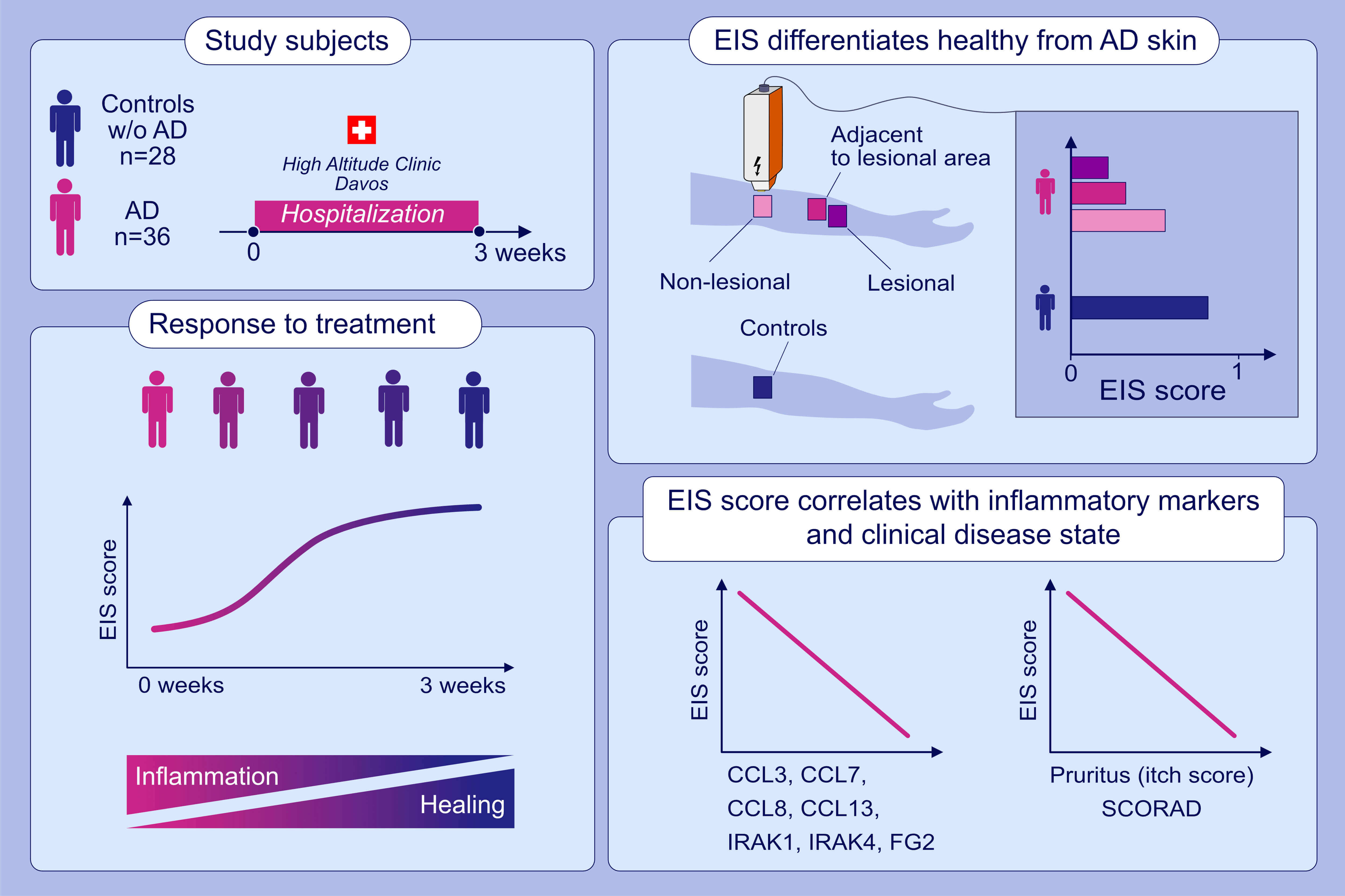
Electrical impedance spectroscopy for the characterization of skin barrier in atopic dermatitis
Arturo O. Rinaldi et al.
Abbreviations: AD, atopic dermatitis; CCL, C-C motif chemokine ligand; CXCL, C-X-C motif chemokine ligand; IRAK, interleukin 1 receptor associated kinase; FG2, fibroblast growth factor 2; EIS, electrical impedance spectroscopy; EIS score, the score given to a measurement by the artificial intelligence model based on EIS measurements